Epigenetics & Nuclear Signaling Antibodies 5
Anti-Acetyl-Histone H3-K23 Antibody (CAB18154)
- SKU:
- CAB18154
- Product Type:
- Antibody
- Reactivity:
- Human
- Reactivity:
- Mouse
- Reactivity:
- Rat
- Host Species:
- Rabbit
- Isotype:
- IgG
- Research Area:
- Epigenetics and Nuclear Signaling
Description
| Antibody Name: | Anti-Acetyl-Histone H3-K23 Antibody |
| Antibody SKU: | CAB18154 |
| Antibody Size: | 20uL, 50uL, 100uL |
| Application: | WB IHC IF |
| Reactivity: | Human, Mouse, Rat, Other (Wide Range) |
| Host Species: | Rabbit |
| Immunogen: | A synthetic peptide of human Acetyl-Histone H3-K23. |
| Application: | WB IHC IF |
| Recommended Dilution: | WB 1:500 - 1:2000 IHC 1:50 - 1:200 IF 1:50 - 1:200 |
| Reactivity: | Human, Mouse, Rat, Other (Wide Range) |
| Positive Samples: | HeLa, NIH/3T3 |
| Immunogen: | A synthetic peptide of human Acetyl-Histone H3-K23. |
| Purification Method: | Affinity purification |
| Storage Buffer: | Store at -20°C. Avoid freeze / thaw cycles. Buffer: PBS with 0.02% sodium azide, 50% glycerol, pH7.3. |
| Isotype: | IgG |
| Sequence: | Email for sequence |
| Gene ID: | 8350 |
| Uniprot: | P68431 |
| Cellular Location: | Chromosome, Nucleus |
| Calculated MW: | 15kDa |
| Observed MW: | 17kDa |
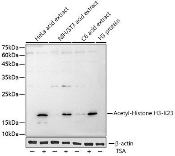
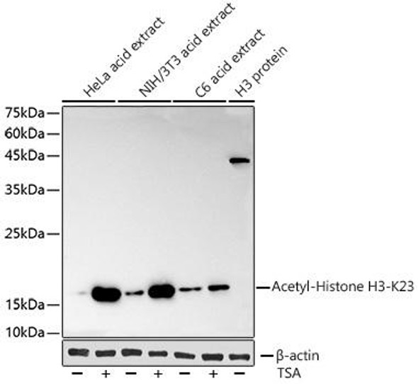
 | Western blot analysis of extracts of various cell lines, using Acetyl-Histone H3-K23 antibody at 1:500 dilution. HeLa cells and NIH/3T3 cells were treated by TSA (1 uM) at 37°C for 18 hours. Secondary antibody: HRP Goat Anti-Rabbit IgG (H+L) at 1:10000 dilution. Lysates/proteins: 25ug per lane. Blocking buffer: 3% nonfat dry milk in TBST. Detection: ECL Basic Kit. Exposure time: 1s. |
 | Immunohistochemistry of paraffin-embedded rat ovary using Acetyl-Histone H3-K23 antibody at dilution of 1:100 (40x lens). Perform microwave antigen retrieval with 10 mM PBS buffer pH 7. 2 before commencing with IHC staining protocol. |
 | Immunohistochemistry of paraffin-embedded human tonsil using Acetyl-Histone H3-K23 antibody at dilution of 1:100 (40x lens). Perform microwave antigen retrieval with 10 mM PBS buffer pH 7. 2 before commencing with IHC staining protocol. |
 | Immunofluorescence analysis of C6 cells using Acetyl-Histone H3-K23 at dilution of 1:100. Blue: DAPI for nuclear staining. C6 cells were treated by TSA (1 uM) at 37°C for 18 hours. Blue: DAPI for nuclear staining. |
 | Immunofluorescence analysis of NIH/3T3 cells using Acetyl-Histone H3-K23 at dilution of 1:100. Blue: DAPI for nuclear staining. NIH/3T3 cells were treated by TSA (1 uM) at 37°C for 18 hours. Blue: DAPI for nuclear staining. |
 | Immunofluorescence analysis of U-2 OS cells using Acetyl-Histone H3-K23 at dilution of 1:100. Blue: DAPI for nuclear staining. U2OS cells were treated by TSA (1 uM) at 37°C for 18 hours. Blue: DAPI for nuclear staining. |
 | Chromatin immunoprecipitation analysis of extracts of HeLa cells, using Acetyl-Histone H3-K23 antibody and rabbit IgG. The amount of immunoprecipitated DNA was checked by quantitative PCR. Histogram was constructed by the ratios of the immunoprecipitated DNA to the input. |


