Description
Human NSFL1C ELISA Kit
| Product Name | Human NSFL1C ELISA Kit |
| Product Code | HUFI03373 |
| Alias | NSFL1C ELISA Kit |
| Detection Method | Sandwich ELISA, Double Antibody |
| Size | 96T |
| Range | |
| Species | Human |
| Storage | 2-8°C for 6 months |
| CV(%) | Intra-Assay: CV <8% Inter-Assay: CV <10% |
| Note | For Research Use Only |
Human NSFL1C ELISA Kit Protocol
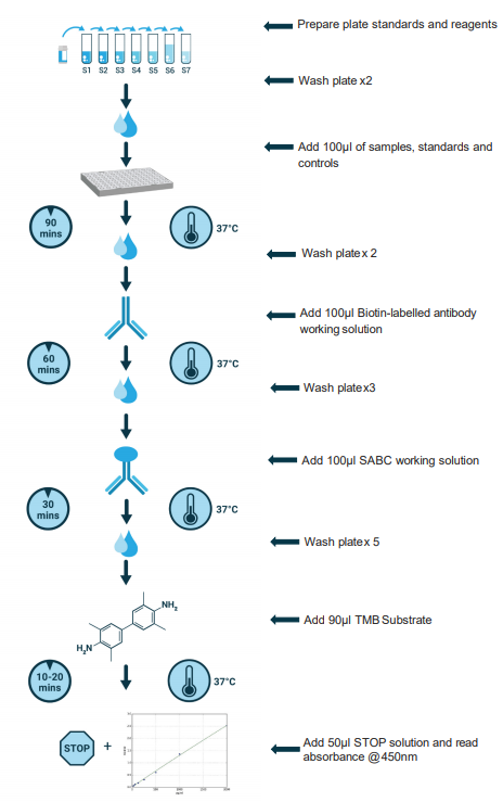
The below protocol is a sample protocol for Human NSFL1C ELISA Kit using a biotinylated detection antibody and streptavidin-HRP. Sandwich ELISAs allow for the detection and quantification of an analyte in a sample by using known analyte concentrations as standards and plotting absorbance of known concentrations vs known standard concentrations. This allows the researcher to calculate the amount of Human NSFL1C present in their sample.
Before adding to wells, equilibrate the SABC working solution and TMB substrate for at least 30 min at 37 °C. When diluting samples and reagents, they must be mixed completely and evenly. It is recommended to plot a standard curve for each test.
Sandwich Protocol

Kit Protocol:
| 1. | Set standard, test sample (diluted at least ½ with Sample Dilution Buffer) and control (zero) wells on the pre-coated plate and record their positions. It is recommended to measure each standard and sample in duplicate. Wash plate twice before adding standard, sample and control (zero) wells! For correct washing procedure, see Page 7, Manual Washing & Automated Washing. |
| 2. | Aliquot 100µl of standard solutions into the standard wells. |
| 3. | Add 100µl of Sample / Standard dilution buffer into the control (zero) well. |
| 4. | Add 100µl of properly diluted sample (serum, plasma, tissue homogenates and other biological fluids.) into test sample wells. |
| 5. | Seal the plate with a cover and incubate at 37 °C for 90 min. |
| 6. | Remove the cover, aspirate the liquid from the plate and wash plate twice with Wash Buffer according to instructions detailed on Page 7. Do not let the wells dry out at anytime. |
| 7. | Add 100µl of Biotin-detection antibody working solution to the bottom of each well (standard, test sample & zero wells) without touching the side walls. |
| 8. | Seal the plate with a cover and incubate at 37°C for 60 min. |
| 9. | Remove the cover, and wash plate 3 times with Wash Buffer. |
| 10. | Add 100µl of SABC working solution into each well, cover the plate and incubate at 37°C for 30 mins. |
| 11. | Remove the cover and wash plate 5 times with Wash Buffer. |
| 12. | Add 90 µl of TMB substrate into each well, cover the plate and incubate at 37°C in dark for 10-20 mins. (Note: This incubation time is for reference only, the optimal time should be determined by the enduser.) As soon as a blue color develops in the first 3-4 wells (with most concentrated standards) and the other wells show no obvious color, terminate the reaction by moving to Step 13. |
| 13. | Add 50µl of Stop solution into each well and mix thoroughly. The color changes into yellow immediately. |
| 14. | Read the O.D. absorbance at 450 nm in a microplate reader immediately after adding the Stop solution. |
Human NSFL1C ELISA Kit components | 96 Assays | Storage |
| ELISA Microplate(Dismountable) | 8×12 strips | 4°C for 6 months |
| Lyophilized Standard | 2 | 4°C/-20°C |
| Sample/Standard Dilution Buffer | 20ml | 4°C |
| Biotin-labeled Antibody(Concentrated) | 120ul | 4°C (Protect from light) |
| Antibody Dilution Buffer | 10ml | 4°C |
| HRP-Streptavidin Conjugate(SABC) | 120ul | 4°C (Protect from light) |
| SABC Dilution Buffer | 10ml | 4°C |
| TMB Substrate | 10ml | 4°C (Protect from light) |
| Stop Solution | 10ml | 4°C |
| Wash Buffer(25X) | 30ml | 4°C |
| Plate Sealer | 5 | - |
Other materials and equipment required:
The Human NSFL1C ELISA Kit will require other equipment and materials to carry out the assay. Please see list below for further details.
- Microplate reader with 450 nm wavelength filter
- Multichannel Pipette, Pipette, microcentrifuge tubes and disposable pipette tips
- Incubator
- Deionized or distilled water
- Absorbent paper
- Buffer reservoir
Sample Preparation
According to best practices, extract protein & perform the experiment as soon as possible after sample collection. Alternatively, store the extracts at the designated temperature (-20°C/-80°C) and for optimal results, avoid repeated freeze-thaw cycles.
| Sample Type | Protocol |
Serum | Use a serum separator tube (SST) and allow samples to clot for 30 mins before centrifugation for 15 mins at ~1000 × g. Remove serum and assay immediately or aliquot and store samples at -20°C or -80°C. |
Plasma | Collect plasma using EDTA or heparin as an anticoagulant. Centrifuge samples at 4°C for 15 mins at 1000 × g within 30 mins of collection. Store samples at -20°C or -80°C. Avoid repeated freeze-thaw cycles. Note: over hemolyzed samples are not suitable for use with this kit. |
Urine & Cerebrospinal Fluid | Collect in a sterile container, centrifuge for 20 mins at 2000-3000 rpm. Remove supernatant and if any precipitation is detected, repeat the centrifugation step. A similar protocol can be used for cerebrospinal fluid. |
Cell culture supernatant | Collect supernatant and centrifuge at 4°C for 20 mins at 2000-3000 rpm. Remove supernatant and rinse cells twice with PBS (pH 7.2-7.4) and perform a total cell count. Optimal cell concentration is 1 million/ml. To release cellular components, dilute the cell pellet in PBS and use 3-4 freeze-thaw cycles in liquid Nitrogen (commercial lyses buffers can be used according to manufacturer’s instructions). Centrifuge at 4°C for 20 mins at 2000-3000 rpm to pellet debris and remove clear supernatant to clean microcentrifuge tube for ELISA or storage. |
Tissue Homogenates | As hemolysis blood can have an impact on assay result, it is necessary to remove residual blood by washing the tissue with pre-cooling PBS buffer (0.01M, pH=7.4).Mince tissue after weighing it and homogenize in PBS (the volume depends on the weight of the tissue. Usually, 9mL PBS would be appropriate for 1 gram tissue pieces. Some protease inhibitors are recommended to add to the PBS) with a glass homogenizer on ice. To further breakdown the cells, you can sonicate the suspension with an ultrasonic cell disrupter or subject it to freeze-thaw cycles. The homogenates are then centrifuged for 5 minutes at 5000×g in order to get the supernatant. The total protein concentration was determined by BCA kit and the total protein concentration of each pore sample should not exceed 0.3mg. |
Fill out our quote form below and a dedicated member of staff will get back to you within one working day!


