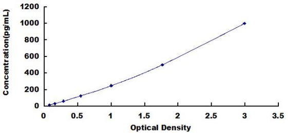
Hamster ELISA Kits
Hamster IL-1b (Interleukin-1b) ELISA Kit
- SKU:
- HMFI0011
- Product Type:
- ELISA Kit
- Size:
- 96 Assays
- Uniprot:
- Q9WVQ9
- Sensitivity:
- 9.375pg/ml
- Range:
- 15.625-1000pg/ml
- ELISA Type:
- Sandwich
- Synonyms:
- IL-1b ELISA Kit, Interleukin-1b ELISA Kit
- Reactivity:
- Hamster
Description
Hamster IL-10 ELISA
IL-1 beta is a cytokine that is mainly produced by macrophages and epithelial cells. IL-1 beta has been shown to play a role in the pathogenesis of IL-1 mediated diseases such as ankylosing spondylitis, osteoarthritis and gouty arthritis. IL-1 beta also plays a role in IL-1 mediated inflammatory diseases such as systemic IL-1 induced erythematosus (SIILE) and IL-1 induced psoriasis. The Assay Genie Hamster IL-1 beta ELISA allows researchers to accurately measure IL-1 beta in hamster serum, blood, plasma, cell culture supernatant and tissue samples.
Key Features
| Save Time | Pre-coated 96 well plate | |
| Quick Start | Kit includes all necessary reagents | |
| Publication Ready | Reproducible and reliable results |
Overview
| Product Name: | Hamster IL-1 beta ELISA |
| Product Code: | HMFI0011 |
| Size: | 96 Assays |
| Target: | Hamster IL-1 beta |
| Alias: | IL-1b, IL-1 beta |
| Reactivity: | Hamster |
| Detection Method: | Sandwich ELISA |
| Sensitivity: | <9.375pg/ml |
| Range: | 15.625-1000pg/ml |
| Storage: | 4°C for 6 months |
| Note: | For Research Use Only |
Additional Information
| Recovery |
| ||||||||||||||||||||
| Linearity: |
| ||||||||||||||||||||
| Intra-Assay: | CV <8% | ||||||||||||||||||||
| Inter-Assay: | CV <10% |
Protocol
*Note: Protocols are specific to each batch/lot. For the correct instructions please follow the protocol included in your kit.
| Step | Procedure |
| 1. | Set standard, test sample and control (zero) wells on the pre-coated plate respectively, and then, record their positions. It is recommended to measure each standard and sample in duplicate. Wash plate 2 times before adding standard, sample and control (zero) wells! |
| 2. | Aliquot 0.1ml standard solutions into the standard wells. |
| 3. | Add 0.1 ml of Sample / Standard dilution buffer into the control (zero) well. |
| 4. | Add 0.1 ml of properly diluted sample ( Human serum, plasma, tissue homogenates and other biological fluids.) into test sample wells. |
| 5. | Seal the plate with a cover and incubate at 37 °C for 90 min. |
| 6. | Remove the cover and discard the plate content, clap the plate on the absorbent filter papers or other absorbent material. Do NOT let the wells completely dry at any time. Wash plate X2. |
| 7. | Add 0.1 ml of Biotin- detection antibody working solution into the above wells (standard, test sample & zero wells). Add the solution at the bottom of each well without touching the side wall. |
| 8. | Seal the plate with a cover and incubate at 37°C for 60 min. |
| 9. | Remove the cover, and wash plate 3 times with Wash buffer. Let wash buffer rest in wells for 1 min between each wash. |
| 10. | Add 0.1 ml of SABC working solution into each well, cover the plate and incubate at 37°C for 30 min. |
| 11. | Remove the cover and wash plate 5 times with Wash buffer, and each time let the wash buffer stay in the wells for 1-2 min. |
| 12. | Add 90 µl of TMB substrate into each well, cover the plate and incubate at 37°C in dark within 10-20 min. (Note: This incubation time is for reference use only, the optimal time should be determined by end user.) And the shades of blue can be seen in the first 3-4 wells (with most concentrated standard solutions), the other wells show no obvious color. |


