APCDD1 Antibody (PACO63443)
")
- SKU:
- PACO63443
")
Description
| Antibody Name: | APCDD1 Antibody (PACO63443) |
| Antibody SKU: | PACO63443 |
| Size: | 50ul |
| Host Species: | Rabbit |
| Tested Applications: | ELISA, WB, IHC |
| Recommended Dilutions: | ELISA:1:2000-1:10000, WB:1:500-1:2000, IHC:1:20-1:200 |
| Species Reactivity: | Human |
| Immunogen: | Recombinant Human Protein APCDD1 protein (22-288AA) |
| Form: | Liquid |
| Storage Buffer: | Preservative: 0.03% Proclin 300 Constituents: 50% Glycerol, 0.01M PBS, pH 7.4 |
| Purification Method: | Antigen Affinity Purified |
| Clonality: | Polyclonal |
| Isotype: | IgG |
| Conjugate: | Non-conjugated |
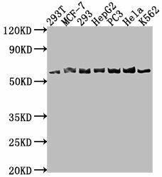 | Western Blot. Positive WB detected in: 293T whole cell lysate, MCF-7 whole cell lysate, 293 whole cell lysate, HepG2 whole cell lysate, PC-3 whole cell lysate, Hela whole cell lysate, K562 whole cell lysate. All lanes: APCDD1 antibody at 1:1000. Secondary. Goat polyclonal to rabbit IgG at 1/50000 dilution. Predicted band size: 59 kDa. Observed band size: 59 kDa. |
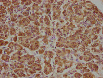 | IHC image of PACO63443 diluted at 1:100 and staining in paraffin-embedded human pancreatic tissue performed on a Leica BondTM system. After dewaxing and hydration, antigen retrieval was mediated by high pressure in a citrate buffer (pH 6.0). Section was blocked with 10% normal goat serum 30min at RT. Then primary antibody (1% BSA) was incubated at 4°C overnight. The primary is detected by a biotinylated secondary antibody and visualized using an HRP conjugated SP system. |
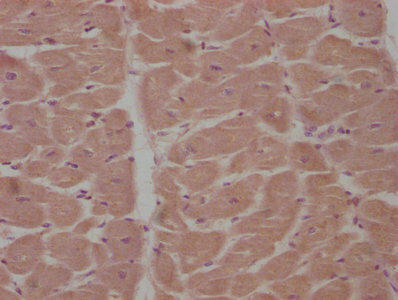 | IHC image of PACO63443 diluted at 1:100 and staining in paraffin-embedded human heart tissue performed on a Leica BondTM system. After dewaxing and hydration, antigen retrieval was mediated by high pressure in a citrate buffer (pH 6.0). Section was blocked with 10% normal goat serum 30min at RT. Then primary antibody (1% BSA) was incubated at 4°C overnight. The primary is detected by a biotinylated secondary antibody and visualized using an HRP conjugated SP system. |
| Background: | Negative regulator of the Wnt signaling pathway. Inhibits Wnt signaling in a cell-autonomous manner and functions upstream of beta-catenin. May act via its interaction with Wnt and LRP proteins. May play a role in colorectal tumorigenesis. |
| Synonyms: | Protein APCDD1 (Adenomatosis polyposis coli down-regulated 1 protein), APCDD1, DRAPC1 |
| UniProt Protein Function: | APCDD1: a single-pass type I membrane protein. Probably plays a role in colorectal tumorigenesis. Directly activated downstream targets of the WNT/beta-catenin pathway. Transcriptional target of beta-catenin/T-cell (Tcf) factor and may enhance cellular proliferation of tumor cells. Down-regulated by wild-type the adenomatosis polyposis coli (APC) protein. Elevated in many primary colon cancer tissues.Protein type: Membrane protein, integralChromosomal Location of Human Ortholog: 18p11.22Cellular Component: integral to plasma membraneMolecular Function: Wnt-protein bindingBiological Process: hair follicle development; negative regulation of Wnt receptor signaling pathwayDisease: Hypotrichosis 1 |
| UniProt Protein Details: | |
| NCBI Summary: | This locus encodes an inhibitor of the Wnt signaling pathway. Mutations at this locus have been associated with hereditary hypotrichosis simplex. Increased expression of this gene may also be associated with colorectal carcinogenesis.[provided by RefSeq, Sep 2010] |
| UniProt Code: | Q8J025 |
| NCBI GenInfo Identifier: | 74728445 |
| NCBI Gene ID: | 147495 |
| NCBI Accession: | Q8J025.1 |
| UniProt Secondary Accession: | Q8J025,Q71M25, B4DUQ0, B4DZT0 |
| UniProt Related Accession: | Q8J025 |
| Molecular Weight: | 58,797 Da |
| NCBI Full Name: | Protein APCDD1 |
| NCBI Synonym Full Names: | APC down-regulated 1 |
| NCBI Official Symbol: | APCDD1 |
| NCBI Official Synonym Symbols: | HHS; HTS; B7323; HYPT1; DRAPC1; FP7019 |
| NCBI Protein Information: | protein APCDD1 |
| UniProt Protein Name: | Protein APCDD1 |
| UniProt Synonym Protein Names: | Adenomatosis polyposis coli down-regulated 1 protein |
| Protein Family: | |
| UniProt Gene Name: | APCDD1 |
| UniProt Entry Name: | APCD1_HUMAN |
Additional Information
Product Type: |
Antibody |
Reactivity: |
Human |
Host Species: |
Rabbit |
Isotype: |
IgG |
Applications: |
ELISA |
Applications: |
WB |
Applications: |
IHC |
Antibody Type: |
Polyclonal Antibody |
Conjugation: |
Unconjugated |
Related Products
")
Anti-APCDD1 Chimeric Recombinant Rabbit Monoclonal Antibody (HDAB0305)
system_update_altDatasheetOverviewSKU:

SPEF2 Antibody
Overview Product Name:SPEF2 AntibodyProduct Code:PACO64977Size:50µLTarget Names:SPEF2Species Reactivity:HumanHos

ompF Antibody
Overview Product Name:ompF AntibodyProduct Code:PACO64865Size:50µLTarget Names:ompFSpecies Reactivity:Escherichia coli

Hmgb1 Antibody
Overview Product Name:Hmgb1 AntibodyProduct Code:PACO64769Size:50µLTarget Names:Hmgb1Species Reactivity:RatHost

RIMS4 Antibody
Overview Product Name:RIMS4 AntibodyProduct Code:PACO64925Size:50µLTarget Names:RIMS4Species Reactivity:HumanHos