Anti-TCF1/TCF7 Antibody (CAB20835)
")
- SKU:
- CAB20835
")
Description
| Antibody Name: | Anti-TCF1/TCF7 Antibody (CAB20835) |
| Antibody SKU: | CAB20835 |
| Antibody Size: | 50µL, 100µL |
| Application: | Western blotting, Immunohistochemistry |
| Reactivity: | Human, Mouse, Rat |
| Host Species: | Rabbit |
| Immunogen: | A synthetic peptide corresponding to a sequence within amino acids 50-150 of human TCF1/TCF7 (NP_003193.2). |
| Application: | Western blotting, Immunohistochemistry |
| Recommended Dilution: | WB 1:500 - 1:1000 IHC 1:50 - 1:200 |
| Reactivity: | Human, Mouse, Rat |
| Positive Samples: | Jurkat, MOLT-4, Mouse thymus, Rat lung |
| Immunogen: | A synthetic peptide corresponding to a sequence within amino acids 50-150 of human TCF1/TCF7 (NP_003193.2). |
| Purification Method: | Affinity purification |
| Storage Buffer: | Store at -20°C. Avoid freeze / thaw cycles. Buffer: PBS with 0.02% sodium azide, 50% glycerol, pH7.3. |
| Isotype: | IgG |
| Sequence: | ELKS SLVN ESEG AAGG AGIP GVPG AGAG ARGE AEAL GREH AAQR LFPD KLPE PLED GLKA PECT SGMY KETV YSAF NLLM HYPP PSGA GQHP QPQP PLHK A |
| Cellular Location: | Nucleus |
| Calculated MW: | 28-42kDa/53-54kDa |
| Observed MW: | 28-50KDa |
| Synonyms: | TCF7, TCF-1 |
| Background: | The protein encoded by this gene is a transcriptional activator that plays an important role in lymphocyte differentiation. This gene is expressed predominantly in T-cells. The encoded protein can bind an enhancer element and activate the CD3E gene, and it also may repress the CTNNB1 and TCF7L2 genes through a feedback mechanism. Several transcript variants encoding different isoforms have been found for this gene. |
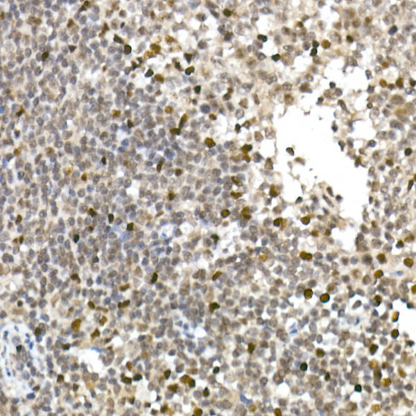 | Immunohistochemistry of paraffin-embedded human tonsil using TCF1/TCF7 Rabbit mAb at dilution of 1:100 (40x lens). Perform high pressure antigen retrieval with 10 mM citrate buffer pH 6. 0 before commencing with IHC staining protocol. |
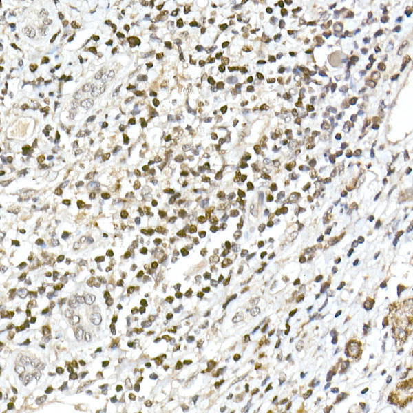 | Immunohistochemistry of paraffin-embedded human liver cancer using TCF1/TCF7 Rabbit mAb at dilution of 1:100 (40x lens). Perform high pressure antigen retrieval with 10 mM citrate buffer pH 6. 0 before commencing with IHC staining protocol. |
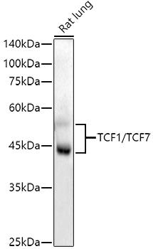 | Western blot analysis of extracts of Rat lung, using TCF1/TCF7 antibody at 1:1000 dilution. Secondary antibody: HRP Goat Anti-Rabbit IgG (H+L) at 1:10000 dilution. Lysates/proteins: 25ug per lane. Blocking buffer: 3% nonfat dry milk in TBST. Detection: ECL Enhanced Kit. Exposure time: 90s. |
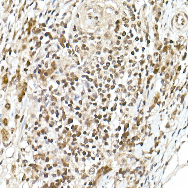 | Immunohistochemistry of paraffin-embedded human colon carcinoma using TCF1/TCF7 Rabbit mAb at dilution of 1:100 (40x lens). Perform high pressure antigen retrieval with 10 mM citrate buffer pH 6. 0 before commencing with IHC staining protocol. |
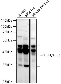 | Western blot analysis of extracts of various cell lines, using TCF1/TCF7 antibody at 1:1000 dilution. Secondary antibody: HRP Goat Anti-Rabbit IgG (H+L) at 1:10000 dilution. Lysates/proteins: 25ug per lane. Blocking buffer: 3% nonfat dry milk in TBST. Detection: ECL Basic Kit. Exposure time: 180s. |
Additional Information
Product Type: |
Antibody |
Antibody Type: |
Secondary Antibody |
Host Species: |
Mouse |
Isotype: |
IgG |
Related Products
")
TCF1/TCF7 Monoclonal Antibody (CAB22458)
TCF1/TCF7 Monoclonal Antibody (CAB22458)The product page for this antibody is being prepared. If you have a question please don't hesitate to get in touch!
")
")
")
")