Anti-PLOD3 Antibody (CAB21066)
")
- SKU:
- CAB21066
")
Description
| Antibody Name: | Anti-PLOD3 Antibody (CAB21066) |
| Antibody SKU: | CAB21066 |
| Antibody Size: | 50µL, 100µL |
| Application: | Western blotting, Immunohistochemistry |
| Reactivity: | Human, Mouse |
| Host Species: | Rabbit |
| Immunogen: | Recombinant fusion protein containing a sequence corresponding to amino acids 25-306 of human PLOD3 (NP_001075.1). |
| Application: | Western blotting, Immunohistochemistry |
| Recommended Dilution: | WB 1:500 - 1:2000 IHC 1:50 - 1:200 |
| Reactivity: | Human, Mouse |
| Positive Samples: | HepG2, MCF7, BxPC-3, Jurkat, Mouse brain |
| Immunogen: | Recombinant fusion protein containing a sequence corresponding to amino acids 25-306 of human PLOD3 (NP_001075.1). |
| Purification Method: | Affinity purification |
| Storage Buffer: | Store at -20°C. Avoid freeze / thaw cycles. Buffer: PBS with 0.02% sodium azide, 50% glycerol, pH7.3. |
| Isotype: | IgG |
| Sequence: | SDRP RGRD PVNP EKLL VITV ATAE TEGY LRFL RSAE FFNY TVRT LGLG EEWR GGDV ARTV GGGQ KVRW LKKE MEKY ADRE DMII MFVD SYDV ILAG SPTE LLKK FVQS GSRL LFSA ESFC WPEW GLAE QYPE VGTG KRFL NSGG FIGF ATTI HQIV RQWK YKDD DDDQ LFYT RLYL DPGL REKL SLNL DHKS RIFQ NLNG ALDE VVLK FDRN RVRI RNVA YDTL PIVV HGNG PTKL QLNY LGNY VPNG WTPE GGCG FCNQ DRRT LPGG QPPP RVFL AVFV EQ |
| Cellular Location: | Lumenal side, Peripheral membrane protein, Rough endoplasmic reticulum membrane |
| Calculated MW: | 84kDa |
| Observed MW: | 85kDa |
| Synonyms: | PLOD3, LH3 |
| Background: | The protein encoded by this gene is a membrane-bound homodimeric enzyme that is localized to the cisternae of the rough endoplasmic reticulum. The enzyme (cofactors iron and ascorbate) catalyzes the hydroxylation of lysyl residues in collagen-like peptides. The resultant hydroxylysyl groups are attachment sites for carbohydrates in collagen and thus are critical for the stability of intermolecular crosslinks. Some patients with Ehlers-Danlos syndrome type VIB have deficiencies in lysyl hydroxylase activity. |
 | Western blot analysis of extracts of Mouse brain, using PLOD3 antibody at 1:1000 dilution. Secondary antibody: HRP Goat Anti-Rabbit IgG (H+L) at 1:10000 dilution. Lysates/proteins: 25ug per lane. Blocking buffer: 3% nonfat dry milk in TBST. Detection: ECL Basic Kit. Exposure time: 180s. |
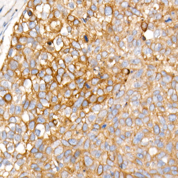 | Immunohistochemistry of paraffin-embedded human esophageal cancer using PLOD3 Rabbit mAb at dilution of 1:1600 (40x lens). Perform high pressure antigen retrieval with 10 mM citrate buffer pH 6. 0 before commencing with IHC staining protocol. |
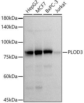 | Western blot analysis of extracts of various cell lines, using PLOD3 antibody at 1:1000 dilution. Secondary antibody: HRP Goat Anti-Rabbit IgG (H+L) at 1:10000 dilution. Lysates/proteins: 25ug per lane. Blocking buffer: 3% nonfat dry milk in TBST. Detection: ECL Basic Kit. Exposure time: 3s. |
Additional Information
Product Type: |
Antibody |
Antibody Type: |
Monoclonal Antibody |
Reactivity: |
Human |
Reactivity: |
Mouse |
Reactivity: |
Rat |
Host Species: |
Rabbit |
Isotype: |
IgG |
Related Products
")
")
PLOD3 Monoclonal Antibody (CAB22354)
OverviewProduct Name:PLOD3 Monoclonal AntibodyProduct Code:CAB22354Reactivity:HumanApplications:Western blotting, Immunohistochem

 ELISA Kit")
Human anti-EPO antibody(anti-Erythropoietin antibody) ELISA Kit
system_update_altDatasheet - तकनीकी मैनुअलsystem_update_altMSDS - तकनीक
")