Anti-IL11RA Antibody (CAB9365)
")
- SKU:
- CAB9365
")
Description
| Antibody Name: | Anti-IL11RA Antibody (CAB9365) |
| Antibody SKU: | CAB9365 |
| Antibody Size: | 50µL, 100µL |
| Application: | Western blotting, Immunohistochemistry, Immunofluorescence |
| Reactivity: | Human, Mouse, Rat |
| Host Species: | Rabbit |
| Immunogen: | A synthesized peptide derived from human IL11RA. |
| Application: | Western blotting, Immunohistochemistry, Immunofluorescence |
| Recommended Dilution: | WB 1:500 - 1:2000 IHC 1:50 - 1:200 IF 1:50 - 1:200 |
| Reactivity: | Human, Mouse, Rat |
| Positive Samples: | K-562, TF-1, Rat brain |
| Immunogen: | A synthesized peptide derived from human IL11RA. |
| Purification Method: | Affinity purification |
| Storage Buffer: | Store at -20°C. Avoid freeze / thaw cycles. Buffer: PBS with 0.02% sodium azide, 50% glycerol, pH7.3. |
| Isotype: | IgG |
| Sequence: | Email for sequence |
| Cellular Location: | Membrane, Single-pass type I membrane protein |
| Calculated MW: | 45kDa |
| Observed MW: | 45KDa |
| Synonyms: | IL11RA, CRSDA, interleukin-11 receptor subunit alpha |
| Background: | Interleukin 11 is a stromal cell-derived cytokine that belongs to a family of pleiotropic and redundant cytokines that use the gp130 transducing subunit in their high affinity receptors. This gene encodes the IL-11 receptor, which is a member of the hematopoietic cytokine receptor family. This particular receptor is very similar to ciliary neurotrophic factor, since both contain an extracellular region with a 2-domain structure composed of an immunoglobulin-like domain and a cytokine receptor-like domain. Multiple alternatively spliced transcript variants have been found for this gene. |
 | Immunofluorescence analysis of TF-1 cells using IL11RA antibody at dilution of 1:100. Blue: DAPI for nuclear staining. |
 | Immunofluorescence analysis of RAW264. 7 cells using IL11RA antibody at dilution of 1:100. Blue: DAPI for nuclear staining. |
 | Immunohistochemistry of paraffin-embedded rat kidney using IL11RA Rabbit mAb at dilution of 1:150 (40x lens). Perform high pressure antigen retrieval with 10 mM citrate buffer pH 6. 0 before commencing with IHC staining protocol. |
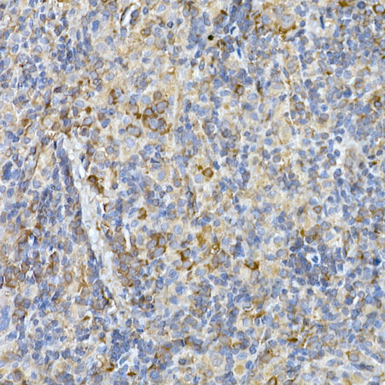 | Immunohistochemistry of paraffin-embedded rat spleen using IL11RA Rabbit mAb at dilution of 1:150 (40x lens). Perform high pressure antigen retrieval with 10 mM citrate buffer pH 6. 0 before commencing with IHC staining protocol. |
 | Immunohistochemistry of paraffin-embedded mouse spinal cord using IL11RA Rabbit mAb at dilution of 1:150 (40x lens). Perform high pressure antigen retrieval with 10 mM citrate buffer pH 6. 0 before commencing with IHC staining protocol. |
 | Immunohistochemistry of paraffin-embedded human lung cancer using IL11RA Rabbit mAb at dilution of 1:150 (40x lens). Perform high pressure antigen retrieval with 10 mM citrate buffer pH 6. 0 before commencing with IHC staining protocol. |
 | Immunohistochemistry of paraffin-embedded human liver using IL11RA Rabbit mAb at dilution of 1:150 (40x lens). Perform high pressure antigen retrieval with 10 mM citrate buffer pH 6. 0 before commencing with IHC staining protocol. |
 | Western blot analysis of extracts of Rat brain, using at 1:1000 dilution. Secondary antibody: HRP Goat Anti-Rabbit IgG (H+L) at 1:10000 dilution. Lysates/proteins: 25ug per lane. Blocking buffer: 3% nonfat dry milk in TBST. Detection: ECL Basic Kit. Exposure time: 180s. |
 | Western blot analysis of extracts of various cell lines, using at 1:1000 dilution. Secondary antibody: HRP Goat Anti-Rabbit IgG (H+L) at 1:10000 dilution. Lysates/proteins: 25ug per lane. Blocking buffer: 3% nonfat dry milk in TBST. Detection: ECL Basic Kit. Exposure time: 30s. |
Additional Information
Product Type: |
Antibody |
Antibody Type: |
Monoclonal Antibody |
Reactivity: |
Human |
Reactivity: |
Mouse |
Reactivity: |
Rat |
Host Species: |
Rabbit |
Isotype: |
IgG |
Related Products
")
Anti-IL11RA Chimeric Recombinant Rabbit Monoclonal Antibody (HDAB0240)
system_update_altDatasheetOverviewSKU:
")
Rat IL11RA Recombinant Protein (RPPB0563)
Cytokines Recombinant Proteins
system_update_altDatasheetsystem_update_altMSDS
")
Human IL11RA/IL11R Alpha Recombinant Protein (RPES4985)
system_update_altDatasheetHuman IL11RA/IL11R Alpha Recombinant Protein Interleukin 11 receptor, alpha subunit (IL11RA/IL-11RA) is a subunit of the interleukin 11 receptor which is a member of the
 ELISA Kit")
Human anti-EPO antibody(anti-Erythropoietin antibody) ELISA Kit
system_update_altDatasheet - तकनीकी मैनुअलsystem_update_altMSDS - तकनीक
")