Anti-HSPA1B Antibody (CAB19317)
")
- SKU:
- CAB19317
")
Description
| Antibody Name: | Anti-HSPA1B Antibody (CAB19317) |
| Antibody SKU: | CAB19317 |
| Antibody Size: | 50µL, 100µL |
| Application: | Western blotting |
| Reactivity: | Human, Mouse |
| Host Species: | Rabbit |
| Immunogen: | Recombinant fusion protein containing a sequence corresponding to amino acids 557-641 of human HSPA1B (NP_005337.2). |
| Application: | Western blotting |
| Recommended Dilution: | WB 1:500 - 1:2000 |
| Reactivity: | Human, Mouse |
| Positive Samples: | MCF7, Mouse testis |
| Immunogen: | Recombinant fusion protein containing a sequence corresponding to amino acids 557-641 of human HSPA1B (NP_005337.2). |
| Purification Method: | Affinity purification |
| Storage Buffer: | Store at -20°C. Avoid freeze / thaw cycles. Buffer: PBS with 0.02% sodium azide, 50% glycerol, pH7.3. |
| Isotype: | IgG |
| Sequence: | GLKG KISE ADKK KVLD KCQE VISW LDAN TLAE KDEF EHKR KELE QVCN PIIS GLYQ GAGG PGPG GFGA QGPK GGSG SGPT IEEV D |
| Cellular Location: | |
| Calculated MW: | 70kDa |
| Observed MW: | 70KDa |
| Synonyms: | HSPA1B, HSP70-1B, HSP70-2, HSP70.2 |
| Background: | This intronless gene encodes a 70kDa heat shock protein which is a member of the heat shock protein 70 family. In conjuction with other heat shock proteins, this protein stabilizes existing proteins against aggregation and mediates the folding of newly translated proteins in the cytosol and in organelles. It is also involved in the ubiquitin-proteasome pathway through interaction with the AU-rich element RNA-binding protein 1. The gene is located in the major histocompatibility complex class III region, in a cluster with two closely related genes which encode similar proteins. |
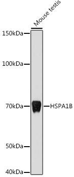 | Western blot analysis of extracts of Mouse testis, using HSPA1B antibody at 1:1000 dilution. Secondary antibody: HRP Goat Anti-Rabbit IgG (H+L) at 1:10000 dilution. Lysates/proteins: 25ug per lane. Blocking buffer: 3% nonfat dry milk in TBST. Detection: ECL Basic Kit. Exposure time: 10s. |
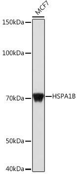 | Western blot analysis of extracts of MCF7 cells, using HSPA1B antibody at 1:1000 dilution. Secondary antibody: HRP Goat Anti-Rabbit IgG (H+L) at 1:10000 dilution. Lysates/proteins: 25ug per lane. Blocking buffer: 3% nonfat dry milk in TBST. Detection: ECL Basic Kit. Exposure time: 1s. |
Additional Information
Product Type: |
Antibody |
Reactivity: |
Human |
Reactivity: |
Mouse |
Host Species: |
Rabbit |
Isotype: |
IgG |
Related Products
")
")

 ELISA Kit")
Human anti-EPO antibody(anti-Erythropoietin antibody) ELISA Kit
system_update_altDatasheet - तकनीकी मैनुअलsystem_update_altMSDS - तकनीक
")